

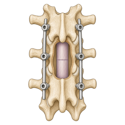
Prepare the repair site
Preserve viable tissue, align the injured ends and establish the mechanical stability that repair requires.

Theseus is developing an AI-guided approach to tissue repair, starting with therapies to reconnect the injured spinal cord.
We develop therapies to reconnect injured tissue. AI models guide treatment design; experiments measure how well function returns.
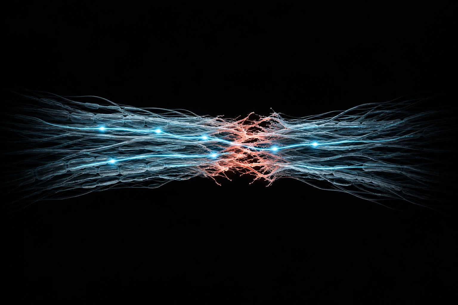 Neural injury
Restore signal continuity.
Our first program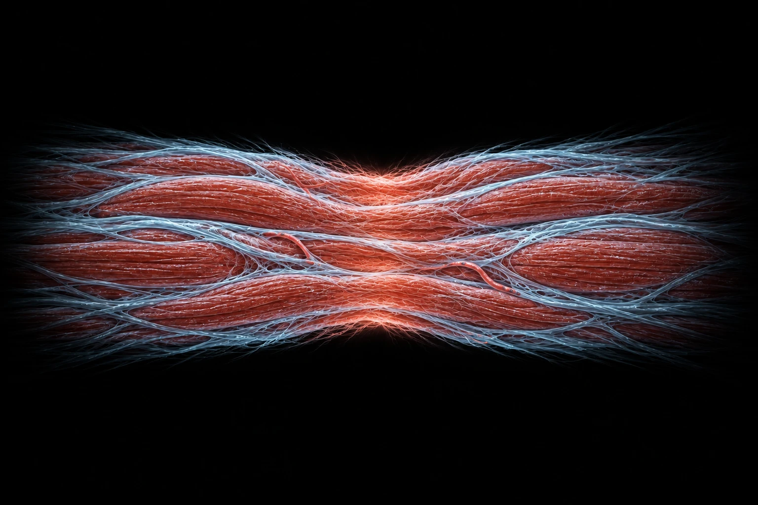 Surgical tissue joins
Rejoin tissue. Prevent leaks.
Research scope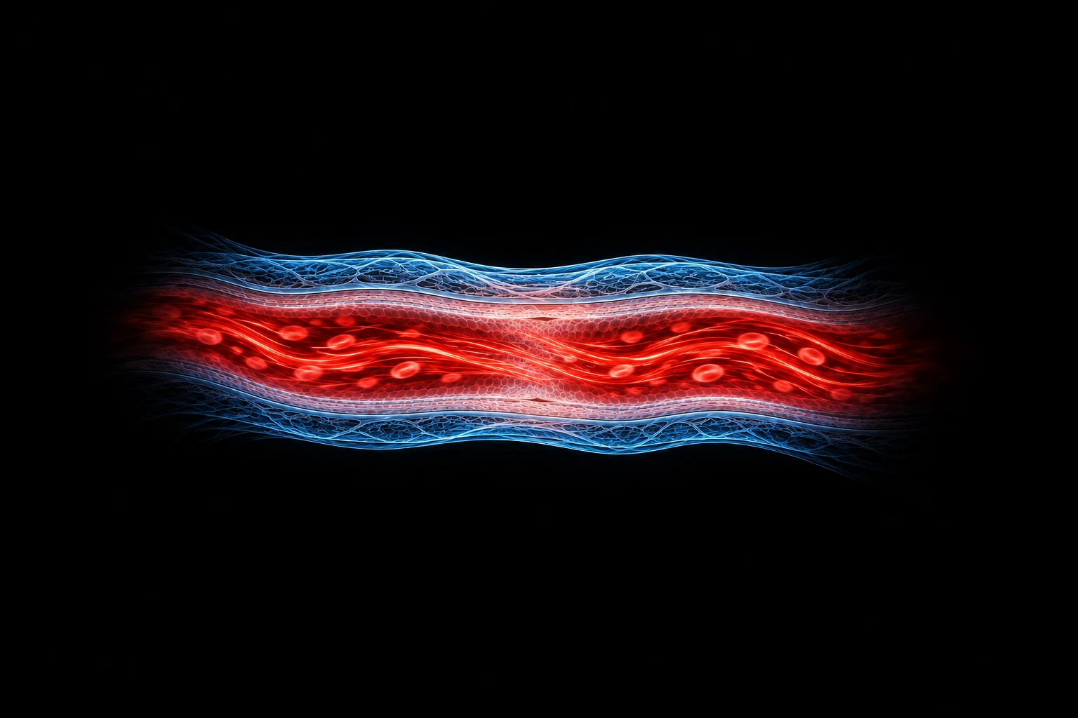 Vascular reconnection
Restore a continuous vessel.
Research scope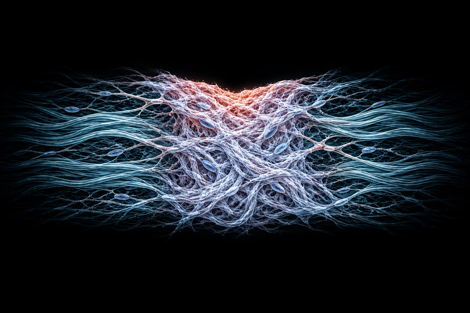 Fibrotic healing
Recover function, not just scar.
Research scope


A formulation alone is not a therapy. We are developing the tissue conditions, fusogen formulation, delivery protocol and recovery pathway as one coordinated approach.
Preserve viable tissue, align the injured ends and establish the mechanical stability that repair requires.
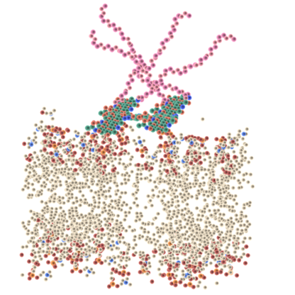
Develop an investigational PEG–chitosan fusogen formulation intended to support membrane repair and neural reconnection.
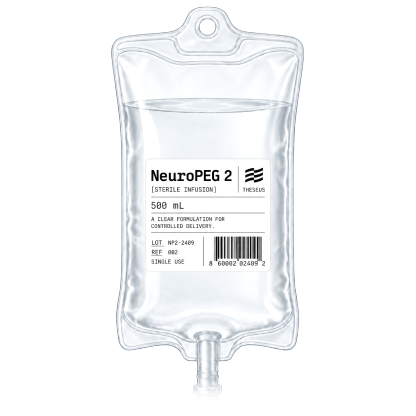
Bring concentration, exposure, timing and treatment sequence into a protocol that can be tested and reproduced.
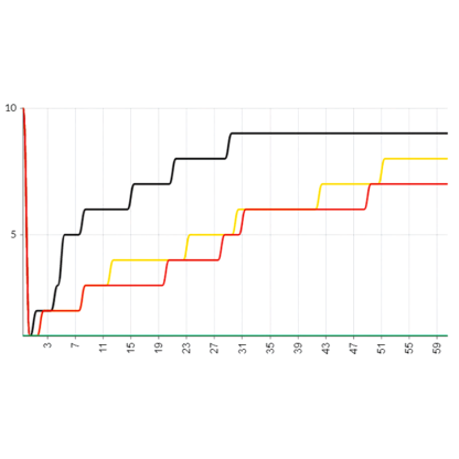
Follow function over time, coordinate rehabilitation and use recovery measurements to refine the whole therapy.
Following complete spinal cord transection, treated animals regained movement in both rabbit and swine studies. The experimental foundation spans rodents, rabbits, large animals and cadaveric procedures.
Movement alone is not enough. Functional scoring, microscopy and axonal tracing cross-check the result at three levels: what the body does, how the tissue connects and where the neural pathways lead.
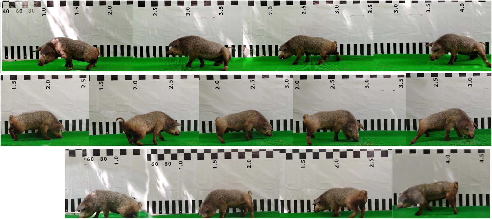
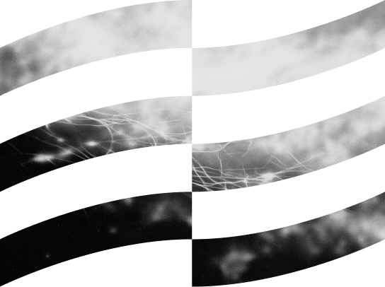
Neurological scores tracked recovery over 60 days. This sequence follows one treated animal walking at days 10, 21 and 40.
Figure 2 · Gait sequence ↗
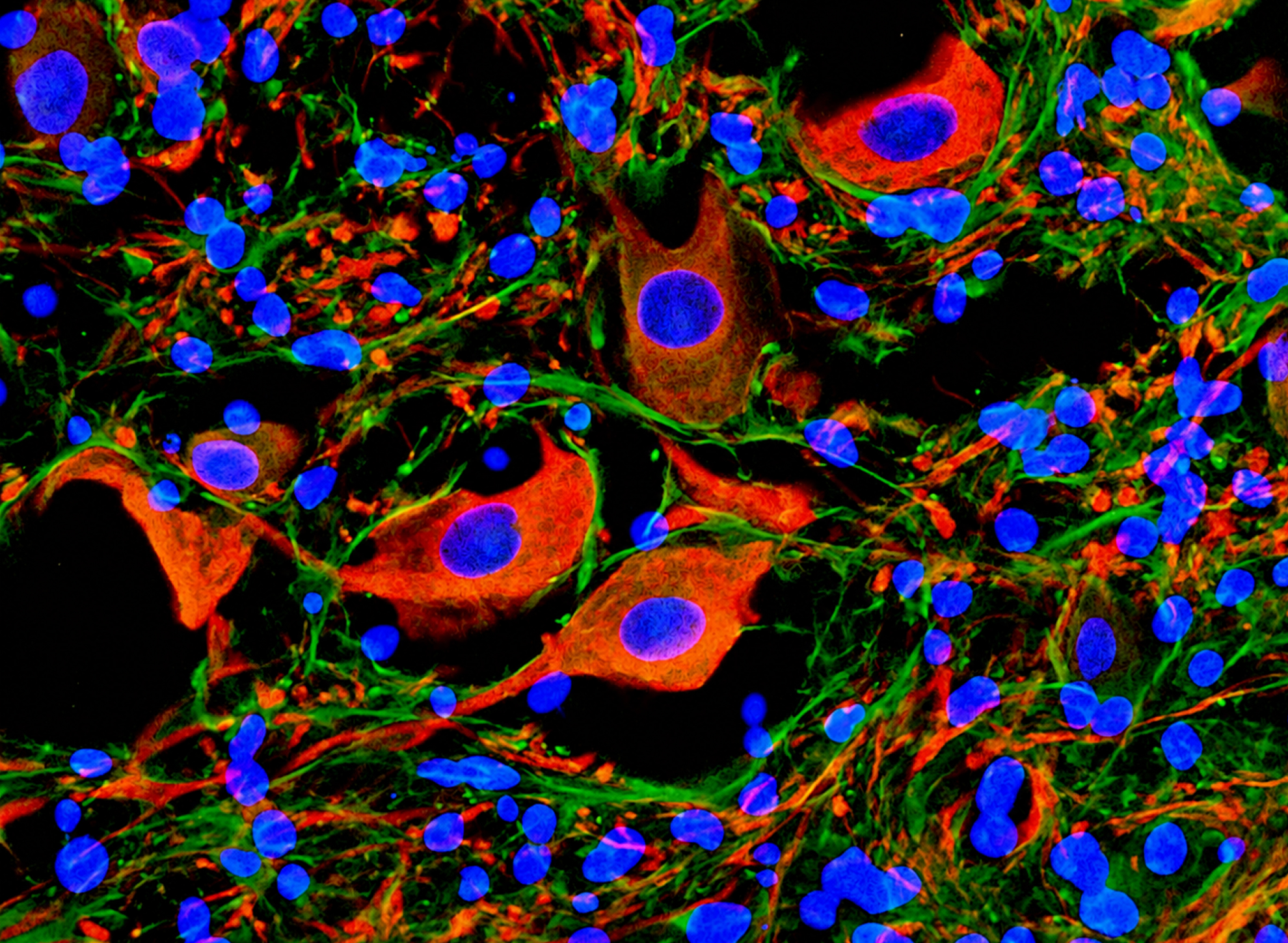
Histology and NF-200 staining reveal tissue continuity and axonal structures at the repair site — a structural check on functional recovery.
Figure 6 · NF-200 / DAPI ↗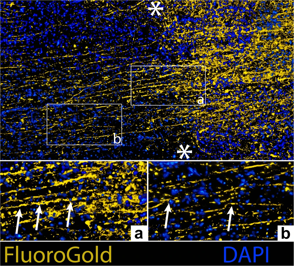
FluoroGold labeling traces neural connections across the lesion. In one treated animal, it provides an anatomical check on reconnection.
Figure 7 · FluoroGold / DAPI ↗Figures 2 & 7: Lebenstein-Gumovski et al., PLOS One, 2026 · CC BY 4.0 · Resized. Microscopy shown as a detail.
Peer-reviewed animal studies in complete-transection models.
Swine: function and structure 3 treated · 2 controls · 60 days Swine: the pilot study 3 treated · 2 controls · 60 days Rabbits: functional recovery 10 treated · 10 controls · 30 daysRandom assignment, blinded neurological scoring, 60-day follow-up. All three treated pigs walked on four limbs; neither control walked independently. Ataxia persisted in treated animals.
Function, tissue morphology and axonal tracing were assessed within the same study. Tracing was performed in one treated animal. These are complementary readouts, not independent replications.
The study evaluated local gel and systemic PEG with surgical stabilization and rehabilitation. These animal results do not establish human efficacy. Clinical contusion injuries require separate validation.
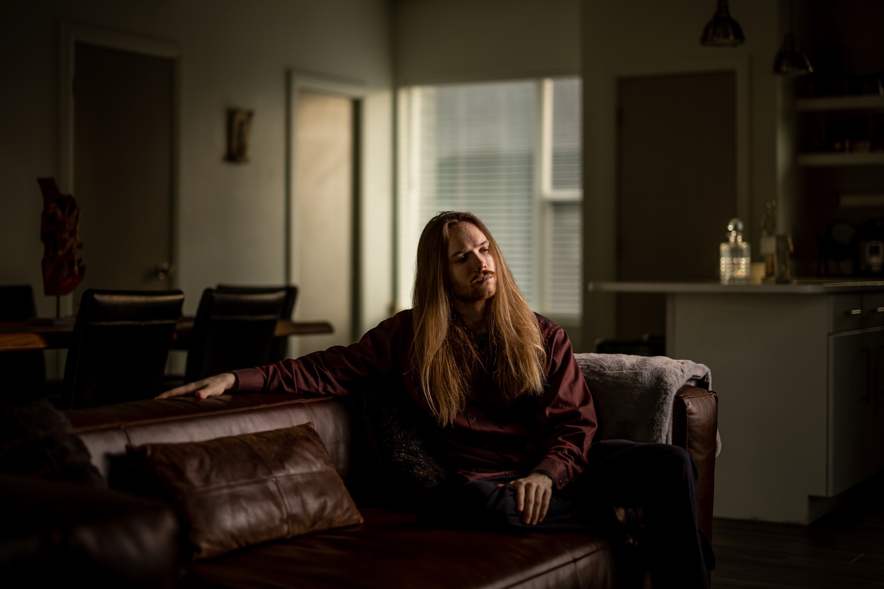
Thiel Fellow, founder of Cryopets, and co-founder of CryoDAO and HydraDAO. He builds organizations advancing cryopreservation, biological replacement and longevity research.
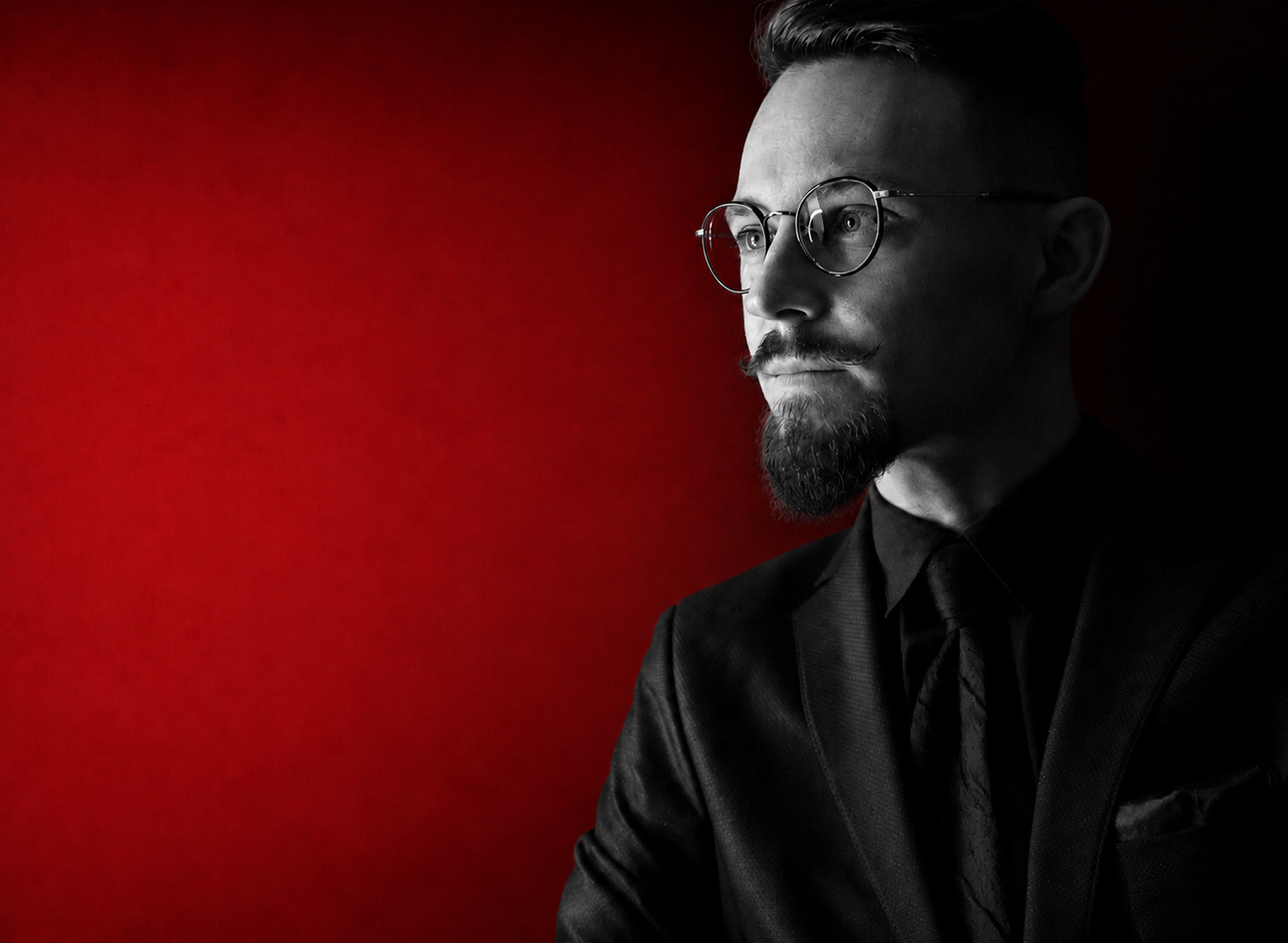
Neurosurgeon and researcher at the Sklifosovsky Institute. He has studied spinal cord injury since 2013, focusing on fusogen-based approaches to restoring neural continuity.
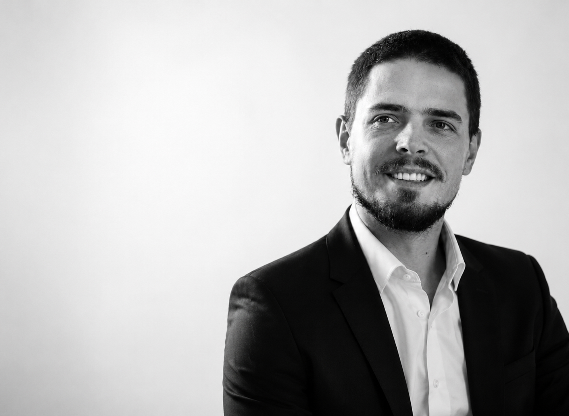
Technology entrepreneur and AI builder. He turns complex, unsolved challenges into mathematical problems that computers can tackle — bringing that approach to the development of biological repair.
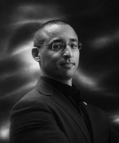
Co-founder of CryoDAO, X-Therma and the Organ Preservation Alliance. He is also a founding ambassador of the American Biostasis Foundation.
Help move tissue repair from an early biological signal toward a reproducible therapy. We welcome scientific collaborators, specialist clinical teams and partners who build for the long term.
Supporting comparative studies and the work toward clinical translation.
Surgical centers and teams to co-develop the operative protocol.
Collaborators extending the preclinical and translational work.
Industry and institutional partners advancing delivery and translation.